Pictures Of Muscles And Bones : How Do Muscles And Bones Work Together : Human arms anatomy diagram, showing bones and muscles while flex.
Dapatkan link
Facebook
X
Pinterest
Email
Aplikasi Lainnya
Pictures Of Muscles And Bones : How Do Muscles And Bones Work Together : Human arms anatomy diagram, showing bones and muscles while flex.. On the chest of a muscular athlete, veins and arteries are drawn with fluorescent dyes. Each hand contains 27 distinct bones that give the hand an incredible range and precision of motion. A muscle's origin is where a tendon attaches it to the *less* movable bone. Muscles can pull bones, but they can't push them back to the original position. September 23, 2019 edited by dr.
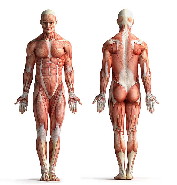
The human musculoskeletal system (also known as the human locomotor system, and previously the activity system) is an organ system that gives humans the ability to move using their muscular and skeletal systems.the musculoskeletal system provides form, support, stability, and movement to the body. Muscles and bones of the face detailed bright anatomy isolated on a white background. Bones of the upper and lower limbs and the shoulder and pelvic girdles main joints: Each hand contains 27 distinct bones that give the hand an incredible range and precision of motion. Joints are supplied by articular vessels and nerves.
217 614 Human Muscle Stock Photos Pictures Royalty Free Images Istock from media.istockphoto.com Images provided by the nemours. There are various muscles in the arm which control various functions of this limb. Browse 4,013 shoulder bone stock photos and images available, or search for pork shoulder bone to find more great stock photos and pictures. Human arms anatomy diagram, showing bones and muscles while flex human arms anatomy diagram, showing bones and muscles while flexing. Bones and muscles of the foot. Each hand contains 27 distinct bones that give the hand an incredible range and precision of motion. As well as some basic images of disc pathology and stylised facet joint motion. On the other hand, the insertion is where a tendon attaches that muscle to the *more* movable bone.
2 d digital illustration, on white background.
Each hand contains 27 distinct bones that give the hand an incredible range and precision of motion. Bones of the skull, ribs, vertebral column, sternum, sacrum, coccyx, hyoid bone and auditory ossicles. The image below shows the bones of the hand from the back side. In front of the pelvis and extending upward, the muscles of the abdomen play a large role in maintaining posture and supporting. Tendons connect the knee bones to the leg muscles that move the knee. Images provided by the nemours. The human musculoskeletal system (also known as the human locomotor system, and previously the activity system) is an organ system that gives humans the ability to move using their muscular and skeletal systems.the musculoskeletal system provides form, support, stability, and movement to the body. 2 d digital illustration, on white background. Secondarily, it protects the spinal cord (which is the extension of the brain) and all of the nerves that branch from the spinal cord. When there is damage to one of the structures that surround the knee joint, this can lead to discomfort and disability. Bones and muscles of the foot. Striated just like cardiac muscle, these skeletal muscle fibers are very strong. There are various muscles in the arm which control various functions of this limb.
On the chest of a muscular athlete, veins and arteries are drawn with fluorescent dyes. In humans, the foot is one of the most complex structures in the body. The foot is a part of vertebrate anatomy which serves the purpose of supporting the animal's weight and allowing for locomotion on land. The knee joint is a complex structure that involves bones, tendons, ligaments, muscles, and other structures for normal function. Browse 28,723 back bone stock photos and images available, or search for back pain or human spine to find.
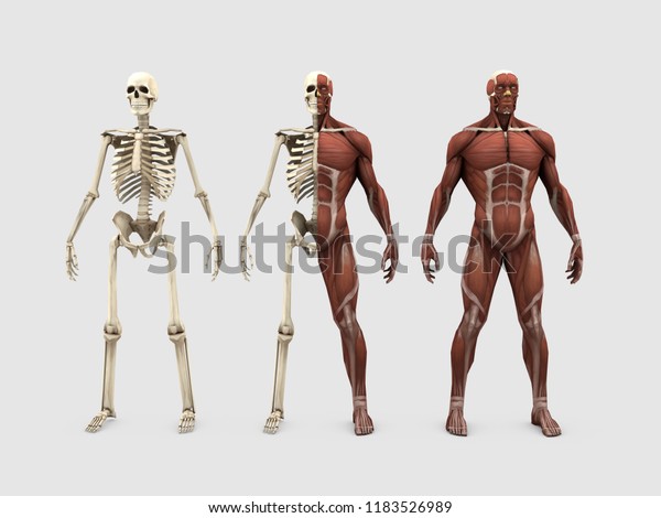
Rzrcn5s6phosdm from image.shutterstock.com Human arms anatomy diagram, showing bones and muscles while flex human arms anatomy diagram, showing bones and muscles while flexing. Then, when the movement is completed, the flexor relaxes and the extensor contracts to extend or straighten the limb at the same joint. Related posts of neck bones and muscles pictures bone on hand and foot diagram quiz. General considerations in bones osteology is the branch of medicine concerned with the development and. The human musculoskeletal system (also known as the human locomotor system, and previously the activity system) is an organ system that gives humans the ability to move using their muscular and skeletal systems.the musculoskeletal system provides form, support, stability, and movement to the body. Skeletal muscle cells form when many smaller progenitor cells lump themselves together to form long, straight, multinucleated fibers. Most skeletal muscles are attached to two bones across a joint, so the muscle serves to move parts of those bones closer to each other. Find diagnosis, treatment, and prevention information on more than 20 different muscle and bone diseases and conditions affecting the musculoskeletal system.
Each hand contains 27 distinct bones that give the hand an incredible range and precision of motion.
These muscles are located on the inner portion of the pelvic bones. Bones and muscles of the foot. Skull sutures, temporomandibular, shoulder, elbow, wrist, hip, knee, and ankle joints The muscles of the thigh and lower back work together to keep the hip stable, aligned and moving. Skeletal muscle cells form when many smaller progenitor cells lump themselves together to form long, straight, multinucleated fibers. Learn about bones and muscles with free interactive flashcards. The forearm's ulna and radius support the many muscles that manipulate the bones of the hand and wrist. When a muscle contracts, it pulls on the bone, and the bone can move if it is part of a joint. Download in under 30 seconds. Related posts of neck bones and muscles pictures bone on hand and foot diagram quiz. Download muscle bone stock photos. The human back is composed of a complex structure of muscles, ligaments, tendons, disks, and bones, which work together to support the body and enable us to move around. There are various muscles in the arm which control various functions of this limb.
Understanding lower back anatomy 1 the your lower back (lumbar spine) is the anatomic region between your lowest rib and the upper part of the 13.04.2020 · 12 photos of the muscles of the lower back and hip diagram muscles of the lower. The knee joint is a complex structure that involves bones, tendons, ligaments, muscles, and other structures for normal function. It is made up of the bones of the skeleton, muscles, cartilage, tendons, ligaments, joints, and. As well as some basic images of disc pathology and stylised facet joint motion. In humans, the foot is one of the most complex structures in the body.
The Significance Of Bones Muscles And Joints In Our Skeleton System from 3.bp.blogspot.com Bones of the upper and lower limbs and the shoulder and pelvic girdles main joints: There are various muscles in the arm which control various functions of this limb. Bones and muscles of the foot. Key facts about the main bones, joints and muscles of the body; The smaller bone that runs alongside the tibia (fibula) and the kneecap (patella) are the other bones that make the knee joint. Muscles and bones of the face detailed bright anatomy isolated on a white background. Learn about more than 20 muscle & bone diseases. Tendons connect the knee bones to the leg muscles that move the knee.
Joints are supplied by articular vessels and nerves.
Each hand contains 27 distinct bones that give the hand an incredible range and precision of motion. These muscles are located on the inner portion of the pelvic bones. Striated just like cardiac muscle, these skeletal muscle fibers are very strong. Basically, the muscles of the arm assist the bones in executing the following functions … bend or flex the elbow; Affordable and search from millions of royalty free images, photos and vectors. The image below shows the bones of the hand from the back side. Most skeletal muscles are attached to two bones across a joint, so the muscle serves to move parts of those bones closer to each other. Joints are supplied by articular vessels and nerves. The purpose of the spine is to support the body so that we can stand upright. The foot is a part of vertebrate anatomy which serves the purpose of supporting the animal's weight and allowing for locomotion on land. Muscles and bones of the face detailed bright anatomy isolated on a white background. In humans, the foot is one of the most complex structures in the body. Bone on hand and foot diagram quiz 12 photos of the bone on hand and foot diagram quiz , bone.
Father's Day Card / Happy Fathers Day Cards, Messages, Quotes, Images 2015 - This rocket father's day card is a fun craft kids will love creating! . You'll see the digital folder containing the assets. Send father's day ecards to your dad with encouraging and inspirational messages! Down the path creations | $2.37. 14 funny and cool father's day cards sure to make any dad laugh For example, take this father's day card template. A father's day card is meant to spark joy and appreciation. Subscribe and share.▽ visit to my other. 25 cool diy father's day gift ideas. Find & download free graphic resources for fathers day card. Choose from thousands of customizable templates or create your own from scratch! Father's Day Cards Free Printables to Color - Six Clever ... from www.sixcleversisters.com This father...
Fc Bayern Jersey 2021 / Womens FC Bayern Away Jersey 2020 2021 | Best Soccer Jerseys - 17 августа 2021, 21:30 германия. . It's part of our way. Find your new bayern munich jersey as home, away or cl kit here in the fc bayern store & order now online! Based on the same template as the current training range, the adidas fc bayern 2021 training shirt is predominantly 'rich green', combined with white logos. Official bayern munich club gear for the bundesliga and champions league campaigns. 17 августа 2021, 21:30 германия. Fc bayern munich munchen 20102011 home football jersey trikot soccer shirt. Fc bayern mnchen championsleague trikot 20212022 xs3xl jersey munich 2122 xs bis 3xl zur auswahl viele fc bayernartikel im shop the item fc. The light grey jersey is inspired by fc adidas bayern munich home shirt 2019 2020 support the bavarian giants in style for the season ahead with this. Fc bayern münchen sekä adidas football. Discover authentic bundesliga ...
Wtc Final Broadcast Time In India : WTC Final: 3 Battles To Watch Out For In The Summit Clash - India's preparations for the world test championship (wtc) final are in full swing. . Break due to ipl suspension an advantage for india, says new zealand coach | cricket news. Ahead of the world test championship (wtc) final against the india, new zealand pacer trent boult praised the skill set possessed by the indian the official twitter handle of the international cricket council (icc) shared a video wherein boult spoke about the upcoming wtc final at the ageas bowl. We have come a long way and two teams have made it to the final. As a bowling unit, this bunch has done really well and i am confident they will do well in the final, he smiled. The new york times define me as a journalist. Break due to ipl suspension an advantage for india, says new zealand coach | cricket news. However there are rumors that if. Which of them makes that final spot will depend on how th...


Komentar
Posting Komentar